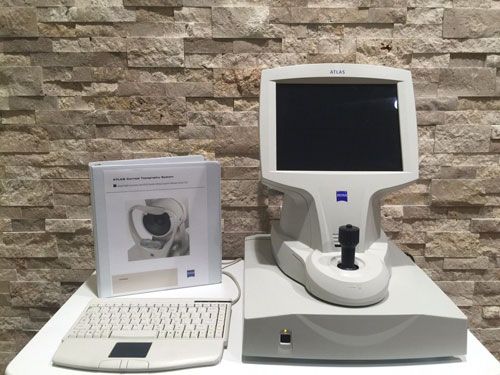
ZEISS Atlas 9000
AMERICA North (USA-Canada-Mexico)
The Atlas 9000 is one of the best and most reliable topographers offered by Carl Zeiss. With its patented Cone-of-Focus Alignment System and Arc-Step Algorithm, the Atlas 9000 delivers sub-micron elevation accuracy. Using triangulation, the advanced ATLAS Arc-Step algorithm provides reliable surface reconstruction, 22-ring Placido disk optimized to avoid ring crossover, which means reliable results for a wide range of patients, and long, comfortable 70 mm working distance minimizes focusing error found in “small cone” systems.
Key Features
By merging proven ATLAS Placido disk technology with Visante OCT pachymetry, Visante Omni now provides comprehensive anterior and posterior topography.
Proven Placido Disk Technology with patented Cone-of-Focus™ Alignment System
SmartCapture™ Image Analysis Technology analyzes multiple images during alignment and automatically selects the highest quality image
MasterFit™ II Contact Lens Software helps streamline the fitting of gas permeable (GP) lenses and guides you through difficult and specialty fits
Data compatibility with previous generation ATLAS Corneal Topography Systems to facilitate data management and patient follow up
SmartCapture analyzes 15 digital images per second during alignment and automatically selects the highest quality image
Next generation image processing provides more repeatable, reliable results, even in difficult cases
Less dependence on operator technique means greater efficiency and fewer repeat exams
Corneal Wavefront Analysis takes corneal topography to a new dimension.
Using ray tracing technology, the ATLAS displays higher-order corneal aberrations, providing valuable insight for patient education and treatment planning.
Educate patients about higher-order aberrations and simulate visual acuity
Assess corneal refraction with image simulation and point spread function
Measure corneal spherical aberration with Zernike analysis to optimize the selection of aspheric IOLs
View the effects of individual higher order corneal aberrations
View changes in contrast sensitivity through the modulation transfer function (MTF)
Pupil size, measured at two levels of illumination (scotopic and photopic @700 nm), provides insight into optical zone under varying light conditions
View centration of LASIK or orthokeratology treatment in relation to pupil center to assess treatment effectiveness
Improve multi-focal contact lenses selection by understanding patient’s pupil size at two levels of illumination
Streamline contact lens selection and fitting with automatic Horizontal Visible Iris Diameter (HVID) meCompact, integrated system with powerful computer and analysis software
Unique chin rest positions patient for easy image capture and wide peripheral coverage and automatically detects OD/OS
Non-visible Placido ring illumination is comfortable for even the most light-sensitive patients
Working Distance: 70 mm
Field of View: 17 mm X 14.5 mm
Placido Rings: 22 (18 superiorly, 22 inferiorly)
Illumination Source: Non-visible infrared (950 nm) LED
Optics: Digital CMOS camera with 1280×1024 pixel resolution
Curvature
Measurement Range: 15 to 95 D (3.5 to 22.5 mm)
Accuracy: ± 0.05 D (± 0.01 mm)
Reproducibility: ± 0.10 D (± 0.02 mm)
HVID (white to white)
Measurement Range: 10.0 to 14.0 mm
Resolution: 0.1 mm
Pupillometry
Acquired Images: Scotopic and photopic (700 nm)
Measurement Range: 0.5 to 11.0 mm
Resolution: 0.1 mm
Views
Axial Curvature
Tangential Curvature
Elevation (Best-Fit Sphere)
Irregularity (Best-Fit Ellipsoid)
Videokeratoscopic (Rings, Scotopic, Photopic)
Keratometry
Refractive Power
Mean Curvature
Corneal Wavefront
Image Simulation
Point Spread Function (PSF)
Modulation Transfer Function (MTF)
Presentation Displays
Single View
Overview
OD/OS Comparison
Difference
Trend with Time
Custom
Dimensions/Weight
52 L x 37 W x 50 H (cm)
39 lbs. (17.7 kg)
Electrical: 100-240V~: 50/60Hz, 2-1A

