 1/1
1/1
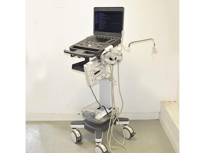
Sonoscape X3
EUROPE (Western and Northern)
SONOSCAPE X3 ULTRASOUND
with
• 6V1 endocavity probe (Acoustic surface: 32 mm x 10 mm,
frequency range from 3 MHz to 15 MHz, image depth: 160 mm)
For ultrasound in gynecology and obstetrics, urology
• 3C-A convex probe from 1 to 6 MHz (Acoustic surface 55 x 14 mm,
frequency range from 1 to 6 MHz, depth 400 mm)
For ultrasound of abdomen, obstetrics, gynecology, urology ,
15" LCD flat screen
Scanning image with extra wide screen in linear range
Connection possibility for external systems with
visualization function prepared on the device
Very large cinema memory
Mains or battery operation for use independent of the electricity network.
Archiving images:
PC connection prepared
Dual USB 2.0
DICOM
Internal image memory on large hard drive
Software :
THI Tissue Harmonic Image (Pulse Inversion Harmonic Image)
Auto – IMT (Intima media) measurement
M-Tuning, optimal image and Doppler adjustments at the push of a button
U-Scan for Spot Reduction
Spatial composite imaging for maximum image harmony
Color Doppler, Triplex, PW and CW
Directional Power Doppler
Trapezoid imaging
Managing Presets
Microscan
Dynamic multi-beam processing technology
Spatial imaging of compounds
Scanning image processing technology
Pure inversion harmonic imaging
Ultimate flexibility
Laptop size and light weight
Magnesium alloy body with 15.6" display
180 degree folding angle
Anti-glare screen and automatic brightness adjustment
Integrated trolley with height adjustment
Silent system
Short startup time
Various accessories
Comfortable backpack and travel bag
WIFI and Bluetooth connection
HDMI