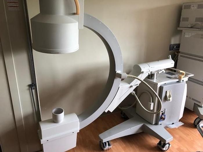
Philips Pulsera BV
AMERICA North (USA-Canada-Mexico)
Dual monitors
12 "Intensifier II
Basic or advanced vascular orthopedic software (optional)
KV / MA range: 0.1 mA at 40 kV to 3 mA at 100 kV.
Focal point: 0.6.
Grid: fixed circular grid 44 lines / cm, ratio 8, SID = 90 cm.
Iris Diaphragm: Electronically controlled iris diaphragm, automatically limited
to the size of the image intensifier input field (15 cm 0).
Remote control and steplessly adjustable to a 5 cm diameter field size
in the image intensifier input field.
Blinds: two parallel shutters of 0.5 cubic mm, remotely controlled
and stepless dimmable for a field of 1 to 16 cm wide in the image intensifier.
The blinds can be rotated ± 90 °; in the intermediate position,
the opening of the slit is perpendicular to the length of the generator cover.
Fluoroscopy: voltage deviation ± 6% ± 2KV, current deviation ± 7% ± 0.02 MA.
X-ray: voltage deviation ± 10%, current deviation ± 10%, deviation ± 3% of time
Includes installation and post-sale training

