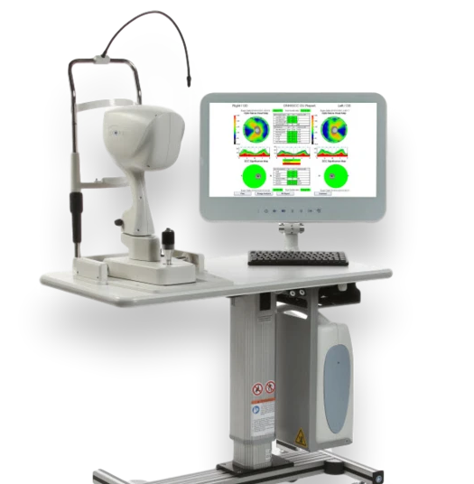
Optovue, ZEISS iVue 2 system WINDOWS 10 with Lens, table
AMERICA North (USA-Canada-Mexico)
WINDOWS 10 INSTALLED
Fully Calibrated and warrantied
Advanced diagnostic imaging capabilities
The iVue® platform supports highly advanced diagnostic imaging applications off the shelf including retina, optic nerve head and anterior segment assessments.
Ganglion Cell Complex (GCC®)
Advanced GCC imaging reveals ganglion cell and axon loss in optic nerve head disease. GCC thickness mapping improves clarity in structural change identification. Optovue’s exclusive FLV% and GLV% analyses increase GCC sensitivity and specificity
3D
3D view provides multi-layer, high-resolution virtual dissection of the retina and optic disc, and depicts them in a way that preserves the retina’s natural curvature. This reduces distortion for simpler interpretation and enhanced 3D visual assessment.
Cornea Advance
Cornea Advance expands iVue’s clinical utility for pre-surgical planning and post-operative assessment. Analysis of the anterior chamber and cornea is enhanced via anterior segment angle measurements and pachymetry with change analysis.
Retina mapping and analysis
Retina mapping with normative comparison, retina change analysis and 3D retinal imaging with en face presentation are all depicted in high-resolution, easy-to interpret color reports.
Optic disc and RNFL assessment
Advanced capabilities include RNFL and GCC® combination reports with normative comparison as well as RNFL and GCC trend analysis —both standard.
Comprehensive anterior segment analysis
Anterior segment capabilities include highly detailed reports for pachymetry mapping with change analysis and anterior segment angle measurement.
Licenses included: 3D, GCC, iWellness Unlimited

