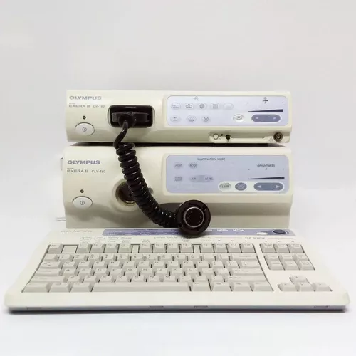
Olympus CV-180 Video Endoscopy Tower
OCEANIA
The Olympus CV-180 complete endoscopy system has
a high-resolution HDTV imaging capability.
This helps ensure the best possible image quality for
endoscopic and laparoscopic procedures.
The CV-180 endoscopy tower is compatible with EVIS 100,
130, 140 Series and EVIS EXERA 160 Series scopes,
bronchoscopes, and surgical endoscopes.
Olympus CV-180 Endoscopy System Includes:
Olympus Video Processor CV180
Olympus Xenon light source CVL 180
Olympus 26” HD monitor MAJ-1462
Olympus MAJ-1933 Keyboard + Link Cable MAJ-1428
Olympus Pigtail Video Cable MAJ-1430
Olympus Water Bottle MAJ-901
Olympus MAJ-1933 Keyboard + Link Cable
Olympus endoscopy trolley with power points.
Accessories included: Biopsy Forceps, Cleaning Brushes, and Foreign Body Retrieval Forceps.
Different scope configurations are available upon request.
Olympus CV-180 Specifications:
General
Brand: Olympus
Model: CV-180
Category: Endoscopy Equipment
Olympus CV-180 Processor
Media: Olympus specifies the XD-Picture Card
(512/256/128/64/32/16 MB). MAPC-10 can be used as a
PC card adapter
Recording Format: TIFF: no compression,
SHQ: approx. 1/3, HQ: approx. 1/5, SQ: approx. 1/10
Number of Recordings: In 16 MB, NTSC/PAL,
TIFF: approx. 17/14 images, SHQ: approx. 80/60 images,
HQ: approx. 200/150 images
Signal Output: HDTV/SDTV
Olympus CVL-180 Lightsource
Light Source: 300-watt xenon lamp
Front Panel: Backlit
Filters: Specially coated filters for NBI (Narrow Band Imaging)
Visualization: Automatically adjusts the light intensity to
achieve ideal illumination for observation
Features:
CV-180
It has high-resolution HDTV imaging capability to provide the
best possible image quality for endoscopes and laparoscopes,
enabling observation of capillaries, mucosal structures,
and other patterns.
Compatible with EVIS 100/130/140 Series and EVIS EXERA 160
Series endoscopes as well as bronchoscopes and
surgical endoscopes (EndoEYE videoscopes,
flexible VISERA video scopes, and 1CCD/3CCD camera heads).
NBI (Narrow Band Imaging) enhances the visibility of capillaries
and other structures on the mucosal surface.
Two types of structure enhancement are available —
the original Type A for observation of
larger mucosal structures with high contrast and the new
Type B for observation of smaller structures, such as capillaries.
Electronic magnification enlarges moving images at the touch
of a button on the scope or the keyboard by 1.2X or 1.5X.
HD/SD SDI output for high-quality video image transfer.
Convenient digital-to-digital recording of both still and
moving images. Still, images are stored on xD
Using remote control switches, cards via PC card adapter and
moving images are stored on digital video recorders via
IEEE 1394 (FireWire, DV, iLINK).
Automatic Iris eliminates the need for switching between Peak
and Average required in conventional manual adjustment.
Picture-in-picture display for any combination of endoscopic,
fluoroscopic, ultrasound, and laparoscopic images and
images from the endoscope position detecting unit.
• Convenient index display for documentation.
Scope ID function for easier endoscopy suite management and
for next-generation system expansion.
CLV-180
Equipped with a specially coated filter for narrowband images
It turns off automatically when the unit has not been in use for
an extended period
Automatically adjusts the intensity of light to achieve the
ideal amount for observation of the gastrointestinal tract
Powerful 300 xenon lamp with vigorous light intensity
All indicators and controls on the front panel are illuminated
from the bottom to improve operability

