 1/1
1/1
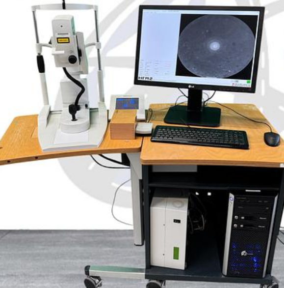
Heidelberg HRA-2 Angiograph
EUROPE (Western and Northern)
Condition Refurbished
1 Introduction1.1 Intended use
The Heidelberg Retina Angiograph 2 is a diagnostic device for
the human eye.Intended use is for the examination of the retina by
laser scanning either with or without contrastagent, resp. fluorescence agent).
The purpose of the angiography testing is to assist in the diagno-sis of eye diseases.
The diagnostic result shall provide help to the physician for adequate therapy.
1.2 The Heidelberg Retina Angiograph 2
The Heidelberg Retina Angiograph 2 is a confocal laser scanning system for digital fluoresceinand indocyanine green angiography.
Fluorescein and indocyanine green angiography can be
per-formed either separately or simultaneously.
For acquisition of the digital confocal images, a laser beam with
a wavelength suitable to excitethe fluorescent dye is focused on the retina.
The laserbeam is deflected periodically by means of oscillatingmirrors to
sequentially scan a two-dimensional sec-tion of the retina.
The intensity of the emitted fluo-rescent light at each point is measured with a
lightsensitive detector. In a confocal optical system, lightemitted outside of
the adjusted focal plane is sup-pressed. This suppression increases with
the distancefrom the focal plane, and results in a high contrastimage.
Furthermore (especially during indocyaninegreen angiography),
the confocal optical system al-lows the user to acquire three-dimensional
imageslayer by layer, which is comparable to laser scanningtomography.
The laser sources contained in the Heidelberg RetinaAngiograph 2 emit laser light of
three different wave-lengths: for fluorescein angiography,
the blue line ofa solid state laser (488 nm) is used to excite the fluo-rescein.
A barrier filter at 500 nm edge wavelengthseparates excitation and fluorescent light.
The samewavelength (but without barrier filter) is used to create red free
reflectance images. For indocya-nine green angiography, a diode laser at
795 nm wavelength is used to excite the dye, while a bar-rier filter at
810 nm edge wavelength separates excitation and fluorescent light.
A second diodelaser at 830 nm wavelength serves to produce infrared
reflectance images.Several image acquisition modes are provided, i.e.
pure fluorescein angiography, pure indocya-nine green angiography,
and simultaneous fluorescein / indocyanine green angiography.
In thesimultaneous mode, each image line is scanned twice.
During the first scan, fluorescein is excitedand its fluorescence is measured;
during the subsequent second scan, indocyanine green is excitedand its
fluorescence is measured. In every acquisition mode, individual images,
time resolved im-age sequences, or a focal series of images
(layered, three-dimensional images) can be acquired.
The images are immediately digitized with a resolution between 384 x 384 and
1536 x 1536 pix-els. The digitized images are stored in
the computer's main memory and then saved on
the com-puter's hard disk.
The size of the field of view can be set to 15 x 15 degrees,
20 x 20 degrees, or30 x 30 degree