GE
AMERICA North (USA-Canada-Mexico)
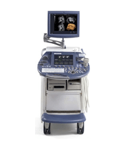
GE Voluson E8
Features
Extraordinary Vision
Next-generation 3D rendering: Dynamic Rendering Engine
Designed for your comfort and efficiency
High resolution transvaginal probe
High resolution flat panel display
Electrical height adjustment
Floating user interface
TruScan architecture
On-board archive including preview and pre-selection
Energy efficient – the Voluson E-Series are among the most energy efficient in the industry.
Speckle Reduction Imaging (SRI) suppresses speckle artifact while maintaining true tissue architecture
CrossXBeam CRI enhances tissue and border differentiation
HD-Flow achieves a more sensitive vascular study and reduces overwriting
New Advanced Volume Contrast Imaging (VCI) with OmniView
VCAD (Volume Computer Aided Diagnosis) – improves workflow by making it easier to acquire volume images of the fetal heart.
System Specifications
Height: 1290 mm (50.8 in) (Adjustable)
Width: 580 mm (22.8 in)
Depth: 930 mm (36.6 in) (Adjustable)
Weight (No Peripherals): 120 kg (265 lb)
Monitor: 19” LCD
Applications
Abdominal
Obstetrical
Gynecological
Small Parts
Vascular/Peripheral
Pediatric and Neonatal
Urological
Cardiology
Neurology
Orthopedic
Transducer Types
Sector Phased Array
Convex Array
Microconvex Array
Linear Array
Volume ‘4D’
Operating Modes
B-Mode (2D)
M-Mode (M)
M-Color-Mode (MC)
Color Flow Mode (C)
Power Doppler Imaging (PDI)
Tissue Doppler Imaging (TD)
HD-Flow Imaging (HD-Flow)
PW Doppler with high PRF (PW)
B-Flow (BF)
Extended View (XTD View)
Coded Contrast Imaging (Contrast Media)
Volume Mode (3D/4D)
Transducers
GE 4C-D – 2-5MHz, Convex
GE M6C – 2-6MHz, Convex
GE SP10-16-D – 7-18MHz, Linear
GE RAB2-5-D – 1-5MHz, Convex Volume
GE RAB4-8-D – 2-8MHz, Convex Volume
GE RIC5-9-D – 4-9MHz, Convex Volume
GE RIC6-12-D – 5-13MHz, Convex Volume
GE RSP6-16-D – 6-18MHz, Linear Volume

