 1/1
1/1
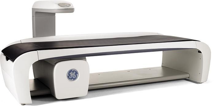
GE Lunar iDXA DEXA
AMERICA North (USA-Canada-Mexico)
The GE Lunar iDXA DEXA was designed to provide you with clinical confidence,
reliability, and productivity for bone health and body composition across all body types,
with research-grade image resolution and clarity with exacting precision.
The Lunar iDXA comes included with automated features that make operation intuitive,
integrated world-class support, and innovative technology.
Whether you’re assessing metabolic health, fracture risk, bone density,
pediatric development or sarcopenia Lunar iDXA gives you a clear glimpse inside
the body.
Benefits:
Clarity
Crisp, high resolution images and precise measurements to help you diagnose
with confidence. Assessment is made easy with Lunar iDXA system's extra-crisp images.
Lunar iDXA offers the latest generation of DXA technology from GE Healthcare.
Its vertebral assessment is comparable to radiographs in identifying
and classifying deformities concerning etiology, grade, and shape,
while using a lower dose of radiation.
Precision
Lunar iDXA's detector technology enables an extremely precise measurement
of the bone, allowing you to track changes that had previously been too minor
to detect. You can better manage treatment plans,
which promotes better patient compliance.
Technology
Lunar iDXA helps with exceptional precision, due to its direct-to-digital detector
plus staggered array and narrow-angle fan beam technology
with Multi-View Image Reconstruction (MVIR).
This corrects magnification error that is observed with competitive wide-angle
fan beam systems.
Versatility
Lunar iDXA is equipped with a wide range of clinical applications
with the enCORE software platform that helps you leverage the best
of the DXA technology for research projects in bone health
or study body composition, i.e. fat and lean mass distribution
and trending for metabolic health applications.
Applications and Features:
BMD: Measures the bone mineral density of a preferred skeletal site
that can be compared to an adult reference population at the sole discretion
of the physician. Generates a reference chart with Z-score and T-score.
AP Spine: Provides an estimate of bone mineral density for the lumbar spine.
Femur/ Dual Femur: Measures both single femur or both the femurs in one scan,
helping you assess the weakest femur through measuring bone mineral density
for the proximal femur.
Color Mapping/ Color Coding: Color Mapping can be used to set thresholds on fat %,
while color coding can be used to code bone, lean tissue and fat tissue.
LVA: Morphometry & Spine Geometry: LVA Morphometry measurement
and analysis provides an x-ray image of the spine for qualitative visual assessment
in order to identify vertebral deformations and estimate vertebral heights
(morphometry), while LVA and APVA Spine Geometry measure Cobb angles.
Dual-energy Vertebral Assessment (DVA): Lateral and anterior views of the spine
with soft tissue equalization to identify vertebral deformities. Performs both LVA
and APVA in one protocol.
Lateral Spine Measurement: Lateral Spine measurement and analysis provides
an estimate of bone mineral density for the lumbar spine.
Hip Axis Length (HAL): Measurement of the distance along the femoral neck axis,
extending from the bone edge at the base of the trochanter to the bone edge
at the inner pelvic brim
Pediatrics: Pediatric measurement and analysis feature provides BMD, BMC,
fat mass, and lean mass for patients from birth to 20 years old.
Atypical Femur Fracture (AFF)*: AFF measurement and analysis provides
an x-ray image of the entire femur for both qualitative visual assessment
and quantitative measures in order to identify areas of focal thickening along
the lateral cortex of the femoral shaft.
FRAX8: FRAX 10-Year Fracture Risk provides an estimate of 10-year probability
of hip fracture and 10-year probability of a major osteoporotic fracture for men
and post-menopausal women ages 40-90 years.
Hand Measurement: Hand measurement and analysis provides an estimate
of the bone mineral density for the hand.
Forearm: Measures radius and ulna, providing additional clinical information on BMD
for the distal forearm. This measurement can be taken in both sitting or supine position.
CoreScan: CoreScan software feature estimates the VAT (Visceral Adipose Tissue)
mass and volume within the android region.
MirrorImage: The MirrorImage function can be used to estimate
the total body composition and bone mineral density (BMD) when regions
of the body are outside of the scan window by using scanned data from
the corresponding region(s) on the opposite half of the body.
One Vision: The OneVision feature allows you to set up multiple measurements
in one exam. This eliminates keystrokes and improves throughput for customers
that routinely perform multiple measurements on each patient.
Trabecular Bone Score (TBS): Provides trabecular bone score based
on bone structure assessment of the trabecular region of the bone.
Small Animal Body Scan: Small Animal measurement and analysis is
for investigational use on laboratory animals or for other tests that
do not involve human subjects.
Orthopedic Hip Implant: Measure the delicate region around the hip implant
and visualizes 19 Gruen zones.
Composer: Composer feature provides many pre-generated report formats
as well as ability to create custom reports.
Quickview: QuickView offers a fast, 10 second spine or femur scan.
Measurement and analysis procedures are the same as other scan modes.
Metabolic Information: Provides insight on metabolic information such
as Resting Metabolic Rate (RMR) and Relative Skeletal Muscle Index (RSMI)
with ability to capture Total Body Water (TBW), Intracellular Water (ICW),
& Extracellular Water (ECW).
Practice Management: Provides general-purpose business reporting tools
to view existing patient population as well as follow- up on next site visit.
Custom Reference Population: Physicians can create a custom reference population
and use that population for comparison to your patients’ results.
OneScan: OneScan performs an AP Spine and Dual Femur exam
without repositioning between scans.
Multi-user Database Access: Allows up to 40 remote computers to be connected
with a common patient database allowing multiple users to access
and analysis patient data.
Body Composition - Total/Regional: Performs total body scan to measure bone mass,
lean mass and fat mass. Also measures regional and whole body bone mineral density
(BMD), lean and fat tissue mass.
Orthopedic Knee: Orthopedic Knee measurement and analysis provides an estimate
of the bone mineral density around knee implants pre and post-surgery.
Pient BMD Trending: Monitoring tool to view changes in a patient’s BMD over time.
To view trending results, all of the trended measurements must be from the same site.
ScanCheck: ScanCheck assists the user in detecting Spine, Femur, Forearm
and Total Body abnormalities.
Sarcopenia*: Sarcopenia software calculates values based on published definitions
and thresholds using measured appendicular lean mass in combination
with patient demographics and entered values of muscle strength
and physical performance.
Narrow Fan Beam Scan: Patented narrow fan beam technology that combines
the best features of pencil beams (no magnification, low dose)
with the short scan time of wide fan beams while reducing magnification error inherent
to wide-angle fan beam systems.
Photon Counting Detector: Dose-efficient photon counting detector technology
that simultaneously counts low and high energy X-rays photons resulting
in lower dosage to the patient and faster and efficient scans.
Lunar iDXA detectors use solid-state crystals (Cadmium Telluride or CdTe)
that absorb the X-ray energy and result in the immediate release of electrons
from their atoms (i.e. direct conversion).
SmartScanTM: Unique feature exclusive to GE Healthcare bone densitometry
systems that identifies bone regions after each transverse sweep to estimate where
to begin exposing the patient to X-rays on the subsequent sweep, thereby reducing
the scan time and the dose to the patient.
K-edge Filter: A unique “K-edge filter” that absorbs the X-rays
in the middle energy range and protects the patient against unnecessary exposure.
Multi-View Image Reconstruction (MVIR): Using narrow fan beam technology
to perform multiple, spaced and transverse sweeps across the site
of interest resulting in accurate determination of bone-height above the tabletop,
minimization of magnification errors, and thereby providing higher precision
and accuracy.
Low Scattered Radiation: Due to narrow-fan beam technology, low scatter radiation
in comparison to wide-angle fan beam systems.
Feature only available with enCORE version 17
Technical Specifications:
Detector technology
CZT-HD Direct-Digital detector (Cadmium Zinc Telluride) in a staggered array
Proprietary design
Patient weight limit
182 kg (400 lbs)
X-ray characteristics
Constant potential power supply (100kV) and K-edge filter (Samarium) for simultaneous Dual-energy X-ray beam.
Radiation
At 1m the operator radiation is at