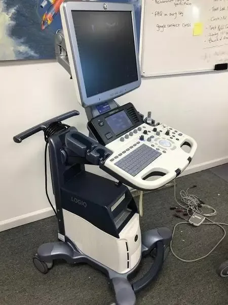
GE Logiq S7 Expert Ultrasound
ASIA (South East)
All items mentioned below including probes, accessories,
options, features and similar properties are solely meant to
provide general information about the
maximum capability of the product.
Some or all features mentioned may not be included in
the listed price. Please contact us directly for
accurate pricing per each additional feature.
Specifications:
Elastography
Flow Quantification
Contrast Imaging
Volume Imaging
SRI-HD (Speckle Reduction)
CrossXBeam Compound Imaging
Scan Assist
Tissue Harmonic Imaging
B-Mode
Doppler
Compatible Ultrasound Probes / Transducers
GE S4-10-D Sector Array Probe
GE L8-18i-D Intraoperative Array Probe
GE RAB4-8-D Curved Array 3D/4D Probe
GE 3SP-D Sector Array Probe
GE C1-5-D Curved Array Probe
GE 11L-D Linear Array Probe
GE 9L-D Linear Array Probe
GE E8C Endocavitary Probe
GE P6D Pencil CW Probe
GE P2D Pencil PW Probe
Next Generation Design With XDclear Imaging
From the start, the GE Logiq S7 Expert Ultrasound has
been designed for the needs of a variety of environments.
Now part of the XDclear family, it includes GE’s premier probe
and platform technology – delivering optimized image
quality across virtually all patient body types.
Multiple applications. Different users. Variety of challenges.
The LOGIQ S7 with XDclear easily handles it, delivering the
high quality images, ease of use,
and specialized capabilities that busy clinicians need.
From its versatility to its budget-friendly price, the LOGIQ S7
with XDclear is designed for practices with high expectations for
both performance and value.
Sensational Performance
Redesigned from top to bottom, the LOGIQ S7 with
XDclear supports productivity and ease in workflow.
S-Agile architecture – Enables images of uniform excellence
across multiple body types with minimal keystrokes
Expanded range of probes – You now get an even wider
selection with both E-Series and XDclear probes,
GE’s highest performing probes. New probes include:
C1-6-D for abdominal exams
C3-10-D for pediatric and neonatal imaging
S2-5-D for cardiac and vascular imaging
Image optimization tools – Automated tools help enhance
image quality such as increased contrast resolution and
improved border definition
Auto TGC – Automatically optimizes image brightness and
contrast, enabling excellent image quality while
streamlining procedures
Smart Design
High-Res widescreen display – High contrast,
configurable 23” LCD monitor with LED backlit display mounted
on an articulating arm allows user to arrange images and
data to meet their needs
10.1-inch touch panel – With a large touch panel, controls are
easy to see and manipulate. Smart keys and
backlit controls further enhance performance and
procedural speed
Engineered for cleanability – The near-seamless exterior has
fewer gaps, making the system easy to clean
Enhanced portability – Small and light-weight, the system
easily navigates crowded exam rooms.
Power Assist battery backup, wireless connectivity and
Bluetooth® printing add to the mobility
Compare Assistant – View a prior study – ultrasound,
mammography, CT or MR – and current images together in
real time via a split screen on the monitor
GE Raw Data – Helps shorten exam times by enabling users to
quickly acquire data and then apply a wide variety of
image processing after the exam
Specialized Capabilities
Well-suited for shared services, the system’s versatility can
be extended with a wide range of clinical packages,
including B-Flow™, Elastography, Vascular Quantification,
B-Steer+, Volume Imaging, and Multi-Modality Imaging Display.
Among the new tools available on LOGIQ S7 with XDclear:
Fetal assessment tools – Advanced STIC
(Spatio-Temporal Image Correlation) helps provide a
high quality, comprehensive fetal echo examination.
Advanced Volume Contrast Imaging (VCI) and
OmniView helps improve contrast resolution and
visualization of rendered anatomy with clarity in
any image plane
Image exchange via smartphone – The ability to
send non-diagnostic images via smartphone and
email is a powerful way to enhance both patient and
clinician satisfaction
Contrast Enhanced Imaging (CEUS) now expanded – Provides a
clear picture of lesion vascularity by optimizing the balance
between penetration and resolution in the liver and can also be
used to enhance the delineation of the LV border in
combination with low MI ultrasound contrast agents.
Features include contrast timer, dual display, timed trigger and
Flash imaging

