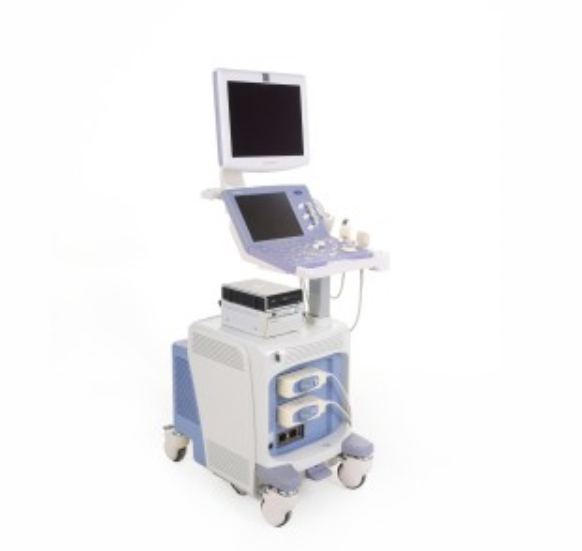
Aloka Prosound Alpha 6
EUROPE (Central and Eastern)
Reconditioned (used)
Technical condition: very good
Visual condition: very good
Real product photos
Japanese production
Power supply: 230 V
Frequency: 50/60 Hz
Power: 900 VA
ALOKA Alpha 6 is a top-class compact ultrasound device using the Aloka Alpha 7
and Alpha 10 system platform, one of the most popular models on the market.
It has a super efficient 12-bit new generation digital converter,
a freely configurable number of transmitting and receiving channels (depending
on the application), and advanced 2nd harmonic technology
(Extended Pure Harmonic Detection - ePHD) directly affects the speed
and reliability of ultrasound examinations. eFlow Doppler imaging mode
(Extended Flow - new generation color Doppler),
combining previously unprecedented sensitivity with simply incredible spatial
and temporal resolution,
Specification:
Compound Pulse Wave Generator - a unique compound wave generator
that controls the amplitude of the generated wave - allows
for extremely precise stimulation of piezoelectric transducers
and frequency control, resulting in an ideal ultrasonic beam
with maximally reduced side lobes and mesh, which directly translates into a sharp
and clear image unattainable with conventional ultrasound systems,
Precise Time Delay Control - precise control of signal delay time,
Dual Focusing - innovative technology of double focusing the beam
in two planes (using classic heads) ensures the highest contrast, spatial
and temporal resolution at a level significantly exceeding previously
known solutions,
Multibeam processing - Multibeam processing offers exceptionally high refresh rates
for optimal performance in dynamic studies,
Super efficient 12-bit digital transducer forming an ultrasonic beam
with a wide dynamic range,
Definitive Tissue Harmonic EchoT (D-THE) - offers clearer edge definition,
reduced sidelobe artifacts and less reverberation noise compared
to fundamental frequency imaging.
Extended Pure Harmonic Detection (ePHD) - additional extended (broadband)
harmonic imaging using the latest achievements in second harmonic imaging -
provides independent detection of phase shift components,
harmonic components and attenuation and backscatter components,
Adaptive Image Processing (AIP) - adaptive image processing.
A fully hardware-implemented function that reduces noise artifacts
and sharpens contours, presenting an ultrasound image in a way similar
to MR imaging,
Spatial Compound Scanning (SCS) - simultaneous scanning of the ultrasonic beam
at many angles, the so-called crossed ultrasound imaging,
Extended Flow (eFlow) - an innovative type of flow imaging (extended flow).
The function has unprecedented resolution and sensitivity, exceeding even
the best Color/Power Doppler imaging. The developed highest spatial
and temporal resolution provides detailed visualization while reducing
the overlap between blood flow and tissue information.
eFlow is an ideal mode for imaging flow in focal lesions or
in the smallest vessels - where Color/Power Doppler imaging is
no longer suitable due to technological limitations.
The function is ideal for imaging the blood supply of suspicious focal lesions
in the breasts, uterus or ovaries - where classic color Doppler
may leave diagnostic doubts - imaging in the eFlow mode dispels these doubts.
Thanks to the highest quality and resolution, eFlow basically allows you
to eliminate time-consuming contrast examinations. EFlow - is
an excellent and fast echocardiographic diagnosis of the fetus
at a level significantly superior to ultrasound systems equipped only
with classic Color Doppler imaging,
Color/Power Doppler - dynamic wide-range new generation Color/Power
Doppler modes provide accurate analysis of blood flow morphology,
Tissue Doppler Imaging (TDI) - Color and Spectral Tissue Doppler that
can present the global distribution of myocardial velocity
and also enables quantitative analyzes such as: velocity profiles,
wall thickness, overload and overload coefficient,
Pulsed Doppler (PW Doppler / PW HPRF Doppler)
and excellent Continuous Doppler (CW Doppler),
Free Angular M-mode (FAM) - M-mode anatomical in real time
and Cineloop memory with 3 cursors (allows you to set cursors
in any position and at any angle). This allows simultaneous display
of 3 M-mode images in different positions in the same time phase,
which facilitates comparison of the peak contraction time in different regions
of the heart,
High Definition Extended Field of View (HDEFV) - precise panoramic imaging
of virtually unlimited length,
eTracking - a unique function enabling early assessment of atherosclerosis
and testing of vascular elasticity. It allows you to automatically track changes
in vessel diameter (with an accuracy of 10 microns) and prepare
a precise pulse wave graph as well as calculate vessel stiffness coefficients.
The test is technically simple, fast, fully automated and repeatable.
eTracking revolutionizes the current approach to the diagnosis
of early atherosclerotic lesions,
Strain / Strain Rate Analysis - rich software for quantitative analysis based
on Tissue Doppler Imaging Analisys,
Kintetic Imaging - kinetic imaging, enables, among others:
automatic endocardial contour and ejection fraction measurement,
A-SMA - software for automatic segmentation quantitative analysis of wall movement,
Dual Dynamic Display (DDD) - simultaneous image display in B-mode + B-mode/Color Doppler or Power Doppler or eFlow in real time,
Quint Frequency Imaging (QFI) allows you to select optimal clinical operating frequencies,
High resolution zoom - allows you to increase the density of lines within the enlarged area,
Image archiving system - a fast, intuitive, easy-to-use system for archiving
and processing ultrasound images and film sequences with a patient database,
reports and comments (over 30,000 patients) allowing for saving images
on a hard drive (HD), flash memory with possibility of data export
and transmission to a computer network compatible with DICOM 3.0,
Wide-angle Transvaginal Imaging - wide-angle transvaginal imaging (180 degrees),
Bi-plane transrectal - possibility of connecting a two-plane rectal head
in the Convex/Convex system (180/180 degrees),
Innovative "Pirouette" chassis ensuring exceptional mobility of the entire system,
Extremely simple and easy to use the camera,
High-class LCD monitor on a movable arm (digital DVI connector),
Excellent ergonomics - colorful, interactive, very large LCD touch panel - 10.4",
Digital outputs: DVI, USB 2.0,
Blue button backlight - new, more economical keyboard layout,
Rich specialized application software,
Freely expandable system architecture,
Imaging modes:
B-Mode,
M-Mode,
Color Doppler,
Power Doppler,
PW Doppler,
eFlow,
Control panel height adjustment in the range of 75-100 cm,
Has normal signs of use,
Menu in English,
Keyboard on the control panel in English,
Included:
User manual in Polish (PDF),
Convex head (UST-9123):
Harmonic imaging (THE – 4 bands, ExPHD – 4 bands),
Operating frequency range: 2.0 to 6.0 Mhz, angle 60 degrees, radius R 60 mm,
Application in: abdominal, obstetric and gynecological examinations,
Linear head (UST-5413):
Harmonic imaging (THE – 4 bands, ExPHD – 4 bands), trapezoidal,
Operating frequency range: from 4.0 to 13.0 Mhz, head length 38 mm,
Application in research: vascular, small organs, musculoskeletal,
Mitsubishi P95 videoprinter,
LG DVD recorder,
Power cord,
Dimensions: 72 x 45 x 155 cm,
Weight: 80 kg,
It has a current inspection and is ready to work,
Issued Technical Passport (Service Report) valid for a period of 12 months,

